Histiocytoma dog
Dog histiocytoma is a small skin mass that is frequently seen in young dogs, mostly under the age of 3.
Although this tumor is quite benign, it is essential to know how to recognize it in order not to confuse it with a malignant lesion that could put your dog's life in danger. Indeed, if the histiocytoma of the dog is not a dangerous lesion, an erroneous health diagnosis can darken the prognosis of the animal by pushing the owner to neglect to take his four-legged friend to a veterinarian when he suffers from a cancerous lesion.
What is histiocytoma in dogs?
The histiocytoma of the dog, sometimes called Langerhansoma, is a small skin mass, more precisely an intradermal nodule, i.e. a relatively spherical, well delimited, mobile and palpable tumor, located on or in the skin of the animal. It is a benign tumor, whose evolution stops naturally after a while and which does not form metastases.
In fact, the histiocytoma usually has a small volume of 1cm to 2cm in diameter on average, and eventually shrinks by itself after reaching its maximum size. This distinguishes it from malignant tumors that, if left untreated, continue to grow uncontrollably and eventually metastasize, invading other organs and causing lethal damage.
Histiocytoma originates from histiocytes (also called Langerhans cells), macrophage cells of the immune system of the body's connective tissue. It should be distinguished from other histiocyte disorders, such as cutaneous or systemic reactive histiocytosis, a chronic disease, and histiocytic sarcoma, a malignant tumor.
Cutaneous histiocytoma in dogs is not contagious or chronic. It is usually a single nodule that appears and disappears on its own without leaving any sequelae. It is, however, a lesion that tends to ulcerate and can become complicated by a local infection if left untreated.
Causes of histiocytoma in dogs
No single cause of histiocytoma in dogs has been identified, but some predisposing factors can be identified. In contrast to most skin tumors in dogs, histiocytomas occur mainly in young animals.
Indeed, about 50% of the cases are diagnosed in dogs younger than 2 years old, and this type of tumor is very exceptional in older dogs. A genetic factor is also suspected since pure breeds are more prone, especially Boxers, Dachshunds, Cockers, English Bulldogs, Labradors, Rottweilers, Spaniels and Schnauzers, to name only the most popular.
There is also a particular form of multiple histiocytoma that can affect different breeds of dogs, but has been described mainly in the Shar Pei. The Bernese Mountain Dog and, to a lesser extent, the Rottweiler and Retrievers (especially the Flat Coated) are predisposed to the development of a malignant histiocyte disease, histiocyte sarcoma.

Symptoms of histiocytoma in dogs
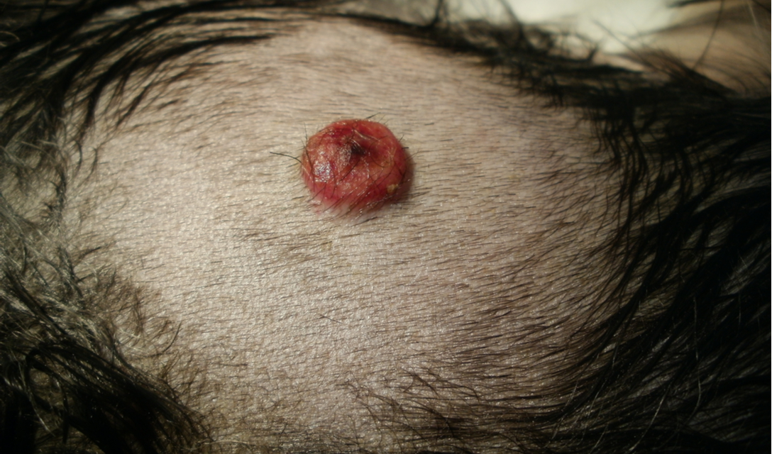
In its most common form, histiocytoma is a single tumor (although multiple forms exist, notably in the Shar Pei), round, smooth, well delimited, easily palpable, found mainly on the front parts of the dog. The surface of this small nodule is most often pink, brilliant and devoid of hairs (alopecic). However, it is common for it to ulcerate, in which case it may become red, visibly irritated and sometimes bloody, crusty and/or purulent.
In some cases, adenopathy (inflammation of the lymph nodes) may accompany the formation of a histiocytoma, but this is not a serious factor - when the diagnosis of a benign mass is confirmed. In the case of a malignant tumor, however, lymph node involvement is a bad sign.
The head of the dog is the preferred site for cutaneous histiocytoma, with more than half of the cases seen in the ears, and a high incidence on the chin and in the region of the lips and muzzle. The neck and front legs of the dog are also regularly affected by this condition, the location of which is very typical and fully involved in the diagnosis.
Although it is more rare, histiocytoma can also affect other parts of the animal's body. The trunk and tail are the other most common sites affected by this condition. In the majority of cases, the size of this small tumor remains small and measures between 1 and 2 cm in diameter.
However, lesions up to 4 cm in diameter are believed to be small nodules only a few millimeters in size. Cutaneous histiocytoma in dogs is painless and usually does not cause any discomfort or suffering to the animal, except in the case of significant ulceration.
Note that ulceration of canine histiocytoma is a common complication. Although it is usually not serious, it requires veterinary treatment. Ulceration of the nodule can further complicate the diagnosis by altering the appearance of the nodule, which may then take on the appearance of a bloody, irregular wound.
Treatment and prognosis of canine histiocytoma
In the majority of cases, it is not necessary to treat canine histiocytoma, as the tumor regresses on its own within a few months. If the lesion does not disappear on its own, surgical removal may be considered, which also allows the diagnosis to be confirmed by laboratory analysis of the removed tissue. If the treatment of this condition is simplistic and the prognosis of the dog is excellent, a scrupulous investigation remains necessary to confirm the diagnosis.
It is essential to ensure that the nodule is benign and not a malignant tumor, which can easily be confused with histiocytoma. Malignant tumors, although they may have a clinical appearance very similar to that of histiocytoma, have a very different prognosis and treatment alternatives.
Additional investigations are particularly indicated in cases of atypical or ulcerated histiocytomas, whose appearance or location do not allow an unambiguous diagnosis by clinical examination alone. Masses located in unusual areas (hind legs, belly, tail, etc.), those with unusual appearance and/or volume (colored, hairy, large, irregular, adherent nodule, etc.), and tumors in dogs older than 3 years of age are particularly suspect.
Furthermore, even in the case of a typical histiocytoma, suspected on clinical examination, further investigation is highly recommended to rule out malignant potential. A malignant tumor should be managed early to avoid a poor prognosis for the dog, and it is not advisable to wait for spontaneous resolution of the lesion - typical of histiocytoma - before making a diagnosis.
In appearance, histiocytoma is relatively similar to mastocytoma, the most common malignancy in dogs. It may also resemble cutaneous lymphoma, cutaneous plasmacytoma, transmissible venereal tumor, and some granulomatous masses.
A cytopunction, a sampling of cells from the nodule, is usually recommended to confirm the diagnosis and/or to prioritize different hypotheses. When a form of multiple histiocytoma is encountered, it is also appropriate to look for cutaneous reactive histiocytosis by histological examination, which will also help to rule out a transmissible venereal tumor.

FAQ
How do I know if my dog has a histiocytoma?
Histiocytoma appears as a small, pink, round, well-defined skin tumor, usually without hair. It is most often a single, small mass (less than 2 cm in diameter), although multiple forms are also seen. Since it can easily be confused with life-threatening malignant skin tumors, veterinary examinations are essential to confirm the diagnosis and provide early management of a possible cancerous lesion.
Can dog histiocytoma be treated?
Yes, it is possible to treat dog histiocytoma by surgical removal, but this is rarely necessary. In most cases, this small, benign nodule will resolve on its own within a few months.
Is dog histiocytoma cancerous?
No, dog histiocytoma is not cancerous. However, it can easily be confused with malignant skin tumors that require early veterinary management to provide the best prognosis for the animal. In particular, mastocytoma and lymphoma, which are common cancerous masses in dogs, may have a similar appearance to histiocytoma.
My dog has a histiocytoma, should I see a veterinarian?
If your dog has what looks like a histiocytoma, it is essential to take him to a veterinarian to have this hypothesis confirmed. It is possible that your dog is suffering from something else entirely, including a life-threatening cancerous tumor.
Dog histiocytoma is not a cancerous skin mass and does not pose any particular danger to your pet's health.
However, it can be easily confused with malignant tumors that need to be treated early to give your pet the best prognosis. Before sighing with relief assuming that your dog has a harmless histiocytoma, it is therefore essential to consult a veterinarian who will reliably confirm this hypothesis.